Anatomy
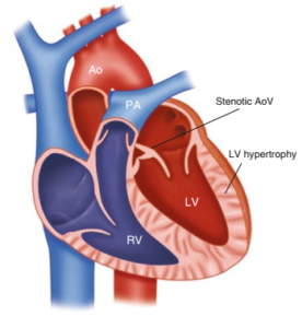
Valvular aortic stenosis
Valvular and vascular stenoses (obstructive lesions) are congenital heart defects (CHDs) in which the only or predominant anatomical abnormality determining the hemodynamics (and consequently the clinical manifestations) is the presence of valvular stenosis—occasionally subvalvular or supravalvular—or stenosis at the level of the great vessels (peripheral pulmonary artery stenosis, coarctation of the aorta). The severity of blood-flow obstruction may vary and may occur at one or more sites within the same or both circulatory systems.
Obstruction of the left ventricular outflow tract (LVOT) may occur at several levels. Valvular aortic stenosis is the most common level of obstruction, while stenosis less commonly involves the other two levels: subaortic (subvalvular) and supravalvular. Left ventricular outflow tract obstruction accounts for approximately 6% of CHDs, with a prevalence of 3–4 per 10,000 live births in the general population.
Aortic valve stenosis (Ao)
The most common anatomical abnormality is a bicuspid aortic valve, which may result in both congenital and acquired aortic stenosis (it represents the anatomical substrate for stenosis developing later in life). Other aortic valve malformations, such as a unicuspid valve and hypoplasia of the aortic annulus, are less common.
Hemodynamics – Common Clinical Manifestations
A. Hemodynamics
The severity of blood-flow obstruction determines the severity of the disease, together with the presence or absence of additional sites of obstruction and other associated anatomical abnormalities (e.g. intracardiac communications).
Isolated obstruction at the level of the outflow valves (aortic or pulmonary) leads to compensatory hypertrophy of the underlying ventricle, enabling it to generate the increased systolic pressure required to maintain adequate blood flow through the stenotic valve.
Cardiac output is maintained at the expense of increased myocardial thickness and the resulting increase in myocardial perfusion requirements (hypertrophy – possible development of ischemia – fibrosis). At the same time, although the hypertrophied myocardium may preserve systolic function, it is characterized by reduced compliance (diastolic dysfunction).
Under conditions of increased cardiac-output requirements, such as exercise, or in the presence of more severe obstruction, cardiac output may become insufficient to meet the body’s demands. Reduced peripheral tissue perfusion also includes reduced perfusion of the myocardium itself, further compromising myocardial blood supply.
Particularly during the vulnerable period of fetal life, reduced cardiac output combined with compensatory myocardial hypertrophy may progress to hypoplasia or atresia of downstream vascular structures and severe myocardial hypertrophy with dysfunction (fibroelastosis), ultimately resulting in hypoplastic left heart syndrome.
B. Clinical manifestations
The severity of the obstruction determines both the degree of compensatory myocardial hypertrophy and the reduction in cardiac output, which in turn determine whether symptoms develop and how severe they are.
Mild stenoses are not associated with symptoms (asymptomatic forms) and may be detected because of abnormal findings on cardiac auscultation (opening sound/click, systolic murmur), other physical findings (reduced intensity of femoral arterial pulses on palpation), or incidentally during an echocardiographic examination performed for another reason.
More severe obstructions, depending on their location (systemic or pulmonary circulation), result in reduced peripheral perfusion and consequently symptoms related to reduced systemic cardiac output (loss of consciousness, syncopal episodes, reduced exercise tolerance, acrocyanosis, centralization of the circulation), reduced pulmonary blood flow (reduced exercise tolerance and/or cyanosis during exercise), reduced myocardial perfusion (angina-like symptoms, precordial pain), or passive pulmonary congestion (pulmonary edema, dyspnea).
Critical obstructions, or combinations of severe obstructive lesions, become apparent immediately during the neonatal period with hypotension and acidosis and may be life-threatening.
Ultrasonography
In mild stenosis, the only ultrasonographic finding may be aliasing on Doppler examination, together with increased flow velocity and, in some cases, post-stenotic dilatation of the aorta. With advancing age, these lesions may progress, and the findings may eventually resemble those of severe stenosis.
Mild aortic valve stenosis with post-stenotic dilatation of the ascending aorta
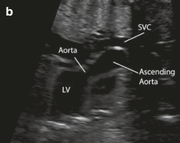
Moderate aortic stenosis with postenotic dilatation
In severe cases, a characteristic second-trimester ultrasonographic finding is a dilated, echogenic left ventricle with impaired systolic function. Mitral valve motion is restricted, with monophasic flow on Doppler and mitral regurgitation. At the foramen ovale, the direction of flow is reversed, from left to right. Pulsed-wave Doppler demonstrates increased blood-flow velocity (>1 m/s).
During the third trimester, persistent obstruction to left ventricular outflow may lead to hypoplastic left heart syndrome, with progressive reduction in the dimensions of the left ventricle.
Treatment
Treatment of valvular aortic stenosis during childhood is primarily interventional (balloon valvuloplasty), since the valve leaflets are relatively flexible and a satisfactory reduction in the pressure gradient can usually be achieved.
Surgical treatment is performed when significant aortic regurgitation is present, either as part of the original disease or as a consequence of balloon valvuloplasty.
Experience to date has shown that balloon valvuloplasty is effective in most cases. Aortic regurgitation following valvuloplasty is observed more frequently in older children (over 11 years of age). The interval before possible future aortic valve replacement tends to be longer when a lower residual pressure gradient is achieved after balloon valvuloplasty and when there is less residual aortic regurgitation.
Patients with aortic stenosis should avoid intense and competitive sports. The risk associated with participation in sports appears to decrease following successful balloon valvuloplasty.
Current recommendations for infective endocarditis prophylaxis recommend antibiotic prophylaxis only in selected high-risk situations, including patients with a previous history of infective endocarditis and those with prosthetic valves. The rationale is that prophylaxis should be directed not simply at patients with an increased risk of developing endocarditis, but particularly at those at greatest risk of serious complications should endocarditis occur.
In selected cases, fetal cardiac catheterization and balloon aortic valvuloplasty may be performed prenatally when ultrasonography demonstrates left ventricular dysfunction, monophasic mitral valve flow, reversal of flow across the foramen ovale, and abnormal/reversed flow through the ductus arteriosus.
Critical aortic stenosis – Neonatal aortic stenosis
Critical aortic stenosis refers to aortic stenosis presenting in the neonatal period that is sufficiently severe for systemic circulation to depend on a patent ductus arteriosus.
When the ductus arteriosus closes, adequate systemic blood flow can no longer be maintained, leading to circulatory shock unless immediate intervention is undertaken.
Prenatal recognition of this potential course is therefore extremely important, as it allows the pregnant woman to be referred and transferred before delivery to a specialized tertiary center where rapid and planned neonatal management can be provided.
Neonates with severe but non-critical aortic stenosis may present with signs of heart failure. The condition is extremely serious and requires management in a specialized referral center.