Anatomy
Coarctation of the aorta (CoA) is a narrowing of the aortic isthmus, typically located distal to the origin of the left subclavian artery and proximal to the insertion of the ductus arteriosus. A related anatomical variant is hypoplasia of the transverse aortic arch, which is also the abnormality most commonly recognized prenatally.
The incidence of isolated coarctation of the aorta is approximately 2 per 10,000 live births, accounting for 7–10% of all congenital heart defects (CHDs).
Clinical Presentation
The clinical presentation can be divided into a neonatal form, which may present either as critical CoA following closure of the ductus arteriosus or somewhat later, usually during the second week of life, and a later childhood form, which typically presents with hypertension or complications related to hypertension.
When the narrowing is extremely severe, approaching atresia, blood supply to the part of the body below the level of the diaphragm may be significantly compromised. In such cases, blood flow to the lower body is maintained through the ductus arteriosus by a right-to-left shunt. Following closure of the ductus arteriosus, the neonate may develop circulatory shock and metabolic acidosis. Immediate intervention is required to reopen or maintain patency of the ductus arteriosus with prostaglandin infusion, followed by transfer to a specialized center for definitive treatment.
When the narrowing is significant but not critical, closure of the ductus arteriosus results in a marked increase in pressure load on the left-sided circulation, potentially leading to left ventricular dysfunction and heart failure. Clinical manifestations usually develop during the first few weeks of life and generally before three months of age.
In older children with CoA, hypertension is frequently detected incidentally during routine examination, often accompanied by a blood pressure difference between the upper and lower extremities. If the diagnosis is missed, CoA may eventually present with complications such as myocardial infarction, stroke, endocarditis, aortic aneurysm and rupture, headaches, epistaxis, intermittent claudication, and a sensation of coldness in the lower extremities.
On physical examination, hypertension with a blood pressure gradient between the upper and lower extremities is typically present. Femoral pulses may be absent or difficult to palpate, and a radiofemoral delay may be detected. On auscultation, a systolic murmur may be heard at the upper left sternal border and in the interscapular region. In longstanding cases, a murmur related to collateral circulation may also be present. Occasionally, an ejection click may be heard in association with a bicuspid aortic valve. Importantly, however, some patients with CoA may have no audible cardiac murmur.
Prenatal Ultrasound
Coarctation of the aorta may be suspected when ventricular disproportion is observed in the four-chamber view, with the right ventricle appearing larger than the left. Except in very severe cases, however, the left ventricle usually continues to form the cardiac apex.
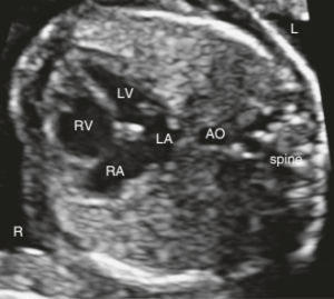
Four-chamber view of the fetal heart showing disproportion between the right and left ventricles.
A similar disproportion may be observed between the great vessels. The pulmonary artery and ductus arteriosus may measure at the upper limits of normal or above normal, whereas the aorta may measure below the lower limits of normal.
Nevertheless, prenatal assessment of these measurements has a significant false-positive rate, because the patent ductus arteriosus modifies fetal hemodynamics and may mask the severity of the lesion.
In contrast to the classic appearance of hypoplastic left heart syndrome, blood flow through both the mitral valve and the aorta is present in CoA. In severe coarctation, left-to-right flow across the foramen ovale may be observed.
Retrograde flow in the aortic arch suggests severe left-sided obstruction and may be associated with aortic valve atresia, severe aortic stenosis, or severe left ventricular hypoplasia.
Particular attention should also be paid to the mitral valve, as a parachute mitral valve may be present, in which the valve leaflets are attached predominantly to a single papillary muscle rather than to two separate papillary muscles.
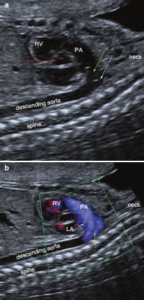
a. Normal aortic arch: the ductus arteriosus (yellow arrow) and the aortic isthmus (white arrow) join the descending aorta.
b. Blood flow through the ductus arteriosus (red arrow) and the aortic isthmus (yellow arrow).
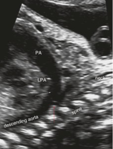
Longitudinal view of the aortic arch showing the ductus arteriosus (white arrow) and the junction of the aortic isthmus with the ductus arteriosus (red arrow), with a smaller diameter and a more perpendicular orientation relative to the descending aorta.
When a ventricular septal defect (VSD) coexists, ventricular disproportion may not be present; instead, disproportion may be limited to the great vessels and may be recognized in the three-vessel and trachea (3VT) view.
In the longitudinal view of the aortic arch, the distance between the left common carotid artery and the left subclavian artery may be greater than normal. At the junction of the aortic isthmus and the ductus arteriosus, an angulation may be observed, producing a characteristic “shelf” appearance.
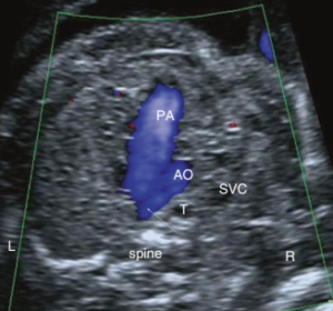
Three-vessel and trachea (3VT) view showing the aorta (Ao) smaller than the pulmonary artery (PA).
During the third trimester, the diagnosis based on ventricular disproportion becomes even more challenging because pathological disproportion may be difficult to distinguish from the physiological right ventricular dominance that normally develops with advancing gestational age.
Management and Clinical Course
CoA should be treated promptly after diagnosis in order to prevent complications associated with hypertension. Without treatment, life expectancy is significantly reduced, historically to approximately 35–40 years, mainly because of cardiovascular complications.
In most centers, the treatment of choice for neonatal CoA is surgical repair, usually involving resection of the narrowed segment followed by anastomosis of the aortic ends.
When significant left ventricular dysfunction is present, stabilization and recovery of ventricular function may be required before definitive surgical repair. During this period, in addition to medical treatment, balloon dilation by cardiac catheterization may occasionally be used as a bridge to improve systemic perfusion and facilitate recovery of left ventricular function.
In older children and adults, cardiac catheterization with balloon angioplasty and stent implantation is an established treatment option and is preferred in many centers for suitable anatomy. Surgical repair is generally favored when long-segment hypoplasia of the aortic arch is present.
In cases of recoarctation, cardiac catheterization with balloon angioplasty, with or without stent implantation, is generally considered the treatment of choice. Catheter-based intervention tends to have a higher likelihood of success when performed on postoperative scar tissue at the site of recoarctation than on native aortic tissue.
Correction should ideally be performed before longstanding systemic hypertension develops. When chronic hypertension has already become established, arterial hypertension may persist even after successful correction of the coarctation, requiring antihypertensive therapy.
Long-term follow-up of patients with CoA includes echocardiography and magnetic resonance imaging (MRI) to detect recoarctation or aortic aneurysm and allow timely intervention. Hypertension should be treated even when it is detected only during exercise.
Patients with an associated bicuspid aortic valve should also be monitored for dilatation or aneurysm of the ascending aorta. When CoA occurs as part of Shone complex, surveillance should also include assessment of the other obstructive lesions affecting the left side of the heart.
Following successful repair and in the absence of significant resting or exercise-induced hypertension, participation in sports is generally permitted. Pregnancy is also usually well tolerated after successful repair, although regular, usually annual, cardiovascular follow-up is recommended.