Physiology of Fetal Circulation
Although the cardiovascular system is described as a single system, both physiologically and anatomically, it can be divided into two circulatory systems: the systemic circulation and the pulmonary circulation. A distinctive feature of fetal circulation is the presence, connection, and support of the placental circulation, which requires significant anatomical and functional adaptations of both the systemic and pulmonary circulations. Understanding these differences, as well as the dramatic changes that occur immediately after birth during the transition from intrauterine to extrauterine circulation, is essential for understanding the timing of presentation of congenital heart defects (CHDs) and the potentially life-saving therapeutic interventions that may need to be undertaken immediately.
Physiology of Circulation After Birth
The systemic and pulmonary circulations are two systems connected in series. Each system consists of a pump (ventricle), vessels that carry blood toward the pump (venous circulation), and vessels that subsequently carry blood away from the pump toward the peripheral tissues (arterial circulation).
Following the direction of blood flow (veins–pump–arteries), the pulmonary circulation consists of the systemic veins, which drain into the superior and inferior vena cava, the right atrium, right ventricle, pulmonary artery and its branches, extending to the pulmonary capillaries.
The systemic circulation consists of the pulmonary veins, which carry blood from the lungs to the left atrium, followed by the left atrium, left ventricle, aorta and its branches, extending to the capillary beds of the body’s organs.
The two circulations are connected at the level of the pulmonary capillaries and the capillary beds of the other organs of the body.
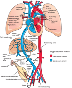
Neonatal circulation (red: oxygenated blood; blue: deoxygenated blood)
The two pumps (ventricles) support two separate circulations that do not normally communicate with each other, and each has different workload requirements. More specifically, the left ventricle must propel blood through an extensive systemic vascular network characterized by relatively high resistance and pressure, whereas the right ventricle pumps blood through the pulmonary circulation, which has lower vascular resistance and pressure. Nevertheless, the output of the two ventricles remains balanced.
Physiology of Fetal Circulation
In fetal circulation, the pulmonary circulation is anatomically present but functionally limited, since oxygenation of fetal blood takes place in the placenta rather than the lungs. Thus, fetal circulation comprises both pulmonary and systemic circulation, while the systemic circulation supplies both the fetal organs and the placental circulation.
Unlike postnatal circulation, the two circulations are arranged functionally in parallel and are interconnected through an intracardiac communication—the foramen ovale—and an extracardiac communication—the ductus arteriosus.
Oxygenated blood from the placenta reaches the fetus through the umbilical vein. Approximately 50% bypasses the liver through the ductus venosus and enters the inferior vena cava, from where it reaches the right atrium. Most of this relatively well-oxygenated blood is preferentially directed through the foramen ovale into the left atrium (right-to-left shunt) and subsequently into the left ventricle.
The remaining approximately 50% of the oxygenated umbilical venous blood perfuses the liver and subsequently reaches the inferior vena cava through the left hepatic vein before entering the right atrium.
From the left ventricle, blood is ejected into the ascending aorta. Approximately 90% supplies the upper part of the fetal body, particularly the brain, while approximately 10% continues through the aortic isthmus into the descending aorta. In this way, the most highly oxygenated blood is preferentially delivered to vital organs, particularly the brain.
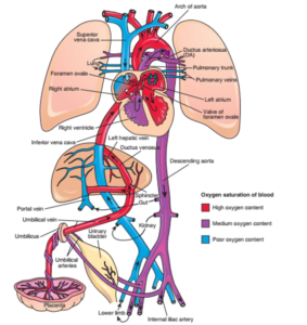
Fetal circulation (red: oxygenated blood; blue: deoxygenated blood)
After oxygen has been delivered to the brain, the now relatively deoxygenated blood returns through the superior vena cava to the right atrium, where it mixes with blood returning through the inferior vena cava. It then passes into the right ventricle and subsequently into the pulmonary artery.
Because fetal pulmonary vascular resistance is high, only approximately 10% of right ventricular output passes through the pulmonary arteries to the lungs, whereas approximately 90% bypasses the lungs through the ductus arteriosus (right-to-left shunt) and enters the descending aorta.
Consequently, the descending aorta contains a mixture of a smaller proportion of relatively oxygenated blood and a larger proportion of less oxygenated blood. This blood either supplies the fetal organs and subsequently returns through the inferior vena cava, or travels through the umbilical arteries and umbilical cord back to the placenta.
At the placenta, fetal blood is oxygenated through gas exchange with the maternal circulation supplied by the uterine arteries, and the circulation then repeats.
Thus, the left ventricle predominantly supplies the brain and upper body, whereas the right ventricle supplies a smaller amount of blood to the lungs and a much larger amount to the lower body and placenta.
Fetal circulation is therefore a parallel, mixed circulation, with right ventricular output approximately 1.4 times greater than left ventricular output.
As pregnancy progresses, changes in placental and pulmonary vascular resistance are accompanied by changes in placental and pulmonary blood flow.
Transition from Fetal to Neonatal Circulation
Immediately after birth, major circulatory changes occur. With separation of the placenta from the circulation, the low-resistance placental vascular bed is removed, resulting in an immediate increase in systemic vascular resistance and systemic arterial pressure.
At the same time, lung expansion and aeration produce a marked reduction in pulmonary vascular resistance and a substantial increase in pulmonary blood flow. Consequently, a greater volume of blood returns through the pulmonary veins to the left atrium.
The resulting increase in left atrial pressure reverses the pressure gradient across the foramen ovale, producing a transient left-to-right tendency and bringing the septum primum into contact with the septum secundum, which together form the valvular mechanism of the foramen ovale. This results in the functional closure of the foramen ovale.
With the onset of pulmonary respiration, arterial oxygen levels increase. The rise in arterial oxygen tension, together with the fall in circulating prostaglandin levels, promotes constriction and subsequent closure of the ductus arteriosus.
The right ventricle therefore ceases to function as part of the systemic circulation: it becomes functionally disconnected from the descending aorta as the ductus arteriosus closes and becomes exclusively connected to the low-pressure pulmonary circulation.