Atrial septal defect (ASD) is one of the most common congenital heart defects (CHDs), accounting for up to 15% of these conditions. It may occur as an isolated defect or in association with other anomalies. The female-to-male ratio is approximately 2:1.
Anatomy
The atrial septum is formed by the incorporation of the septum primum, septum secundum, and structures related to the sinus venosus. During fetal life, the right and left atria communicate through the foramen ovale, which functionally closes after birth as the septum primum comes into close contact with the septum secundum.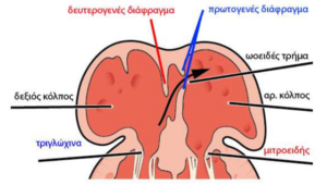
Classification
ASDs are classified into the following types:
• Ostium secundum defect
The defect is located in the central portion of the atrial septum. Its size may range from:
- minimal, as in a patent foramen ovale (PFO)
- to large, as in a significant secundum atrial septal defect
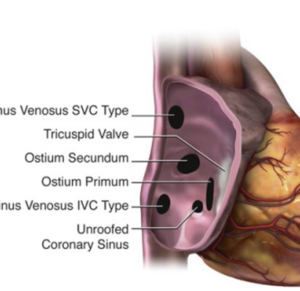
• Ostium primum defect
The defect is located in the inferior part of the atrial septum, immediately above the atrioventricular valves. It is frequently associated with malformation of the mitral valve, particularly a cleft mitral valve. This abnormality belongs to the spectrum of endocardial cushion/atrioventricular septal defects.
• Sinus venosus defect
The defect is located near the junction of the superior or inferior vena cava with the right atrium. Because of its eccentric location, diagnosis may be difficult. It is frequently associated with partial anomalous pulmonary venous connection (PAPVC), in which one or more pulmonary veins drain into the right-sided circulation rather than the left atrium.
• Coronary sinus defect
This rare type is related to partial or complete absence of the wall separating the coronary sinus from the left atrium (unroofed coronary sinus), resulting in an interatrial communication.
ASD is associated with several syndromes, including Holt–Oram syndrome, Noonan syndrome, and Ellis–van Creveld syndrome, as well as chromosomal abnormalities such as trisomy 21 and trisomy 13. Familial forms with autosomal dominant inheritance have also been described and may involve genes such as NKX2-5 and GATA4.
Pathophysiology
During fetal life, an ASD has no significant hemodynamic consequences because right-to-left shunting through the foramen ovale is a normal component of fetal circulation.
After birth, however, an ASD results in a left-to-right shunt at the atrial level. Shunting occurs predominantly during diastole, resulting in volume overload of the right ventricle. Progressive dilation of the right-sided cardiac chambers and increased pulmonary blood flow subsequently develop.
The magnitude of increased pulmonary blood flow, expressed as the Qp:Qs ratio, depends both on the size of the defect and on ventricular compliance. With increasing age, left ventricular compliance decreases, which may result in an increase in the Qp:Qs ratio across the defect.
Cardiac Catheterization
Cardiac catheterization can determine the Qp:Qs ratio and was traditionally used as a reference for deciding whether defect closure was indicated, with a ratio greater than 1.5:1 being used in many reports. It is also the reference method for directly measuring pulmonary artery pressure and pulmonary vascular resistance.
Today, hemodynamic significance can usually be assessed by non-invasive methods, while cardiac catheterization is mainly used therapeutically for transcatheter device closure of suitable defects.
Clinical Course and Management
Patients usually remain asymptomatic, while signs of heart failure may appear during the second or third decade of life.
The development of pulmonary hypertension and Eisenmenger syndrome represents the most serious complication and is associated with significant morbidity and mortality. Pulmonary hypertension has been estimated to occur in approximately 10% of patients and, in exceptional cases, has been reported at a very young age, even around 10 years.
It is generally considered that hemodynamic overload alone may not fully explain the development of pulmonary hypertension and that an individual predisposition may also be involved.
Because correction should ideally be performed before pulmonary hypertension develops, closure, when indicated, is recommended during childhood. If an ASD is diagnosed in a neonate, asymptomatic patients are generally monitored because spontaneous reduction in size or closure may occur, particularly with small secundum defects.
Closure may be performed surgically or, in anatomically suitable secundum ASDs, by transcatheter device closure.
With increasing age, sinus node dysfunction and atrial arrhythmias may develop. Their occurrence appears to be related particularly to the timing of correction and the degree of pre-existing right-sided chamber dilation rather than simply to the method of closure.
Paradoxical embolism may occur in patients with an interatrial communication. However, the presence of an ASD alone does not necessarily constitute an indication for closure for prevention of paradoxical embolism; closure in this setting is recommended only in selected cases according to international guidelines.
Infective endocarditis is extremely rare in an isolated ASD, and routine endocarditis prophylaxis is not recommended.
When surgical treatment is required, it is usually performed during childhood.