Cytomegalovirus (CMV) is a herpesvirus transmitted through sexual contact and infected biological fluids, such as blood, urine, and saliva.
The incubation period ranges from 28 to 60 days (mean: 40 days). In individuals who have not previously been infected, the virus causes viremia lasting approximately 2–3 weeks (primary infection). This is accompanied by the production of immunoglobulin M (IgM) antibodies, followed by immunoglobulin G (IgG) antibodies.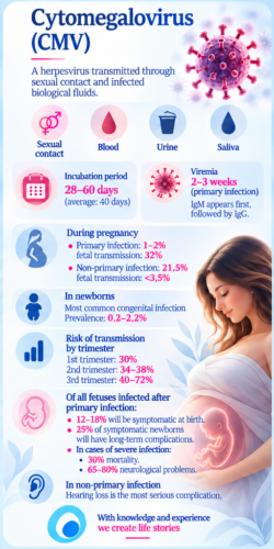
Adults are usually asymptomatic or may develop a mononucleosis-like syndrome characterized by fever, myalgia, malaise, leukocytosis, lymphocytosis, elevated liver enzymes, and lymphadenopathy.
Following primary infection, the virus remains latent in host cells, where it may subsequently reactivate. Reinfection with a different viral strain may also occur (non-primary or secondary infection).
The incidence of primary CMV infection among pregnant women is approximately 1–2%, with a transplacental transmission rate of 32%. Non-primary infection occurs in approximately 21.5% of pregnant women, with a fetal transmission rate of less than 3.5%.
CMV is the most common congenital infection in newborns, with a reported prevalence of 0.2–2.2%.
Transmission may occur through the placenta during pregnancy (the main cause of clinically significant fetal and neonatal complications), through contact with infected cervical secretions during delivery, or through breastfeeding. Infections acquired during delivery or breastfeeding are generally asymptomatic or associated with very mild clinical consequences.
The probability of transplacental transmission increases with advancing gestational age, whereas the severity of clinical manifestations is greater when primary maternal infection occurs earlier in pregnancy.
Transplacental transmission rates by trimester:
First trimester: approximately 30%.
Second trimester: 34–38%.
Third trimester: 40–72%.
Among fetuses infected following primary maternal infection, approximately 12–18% will be symptomatic at birth, and 25% of these will develop long-term complications.
Among infants with severe infection, approximately 30% will die, while 65–80% will develop neurological complications.
In cases of non-primary maternal CMV infection, sensorineural hearing loss is the most serious complication.
Primary Prevention of Cytomegalovirus (CMV) Infection Through Hygiene Measures
Primary prevention aims to reduce the likelihood of exposure to cytomegalovirus (CMV), particularly before conception and during pregnancy.
Seronegative women before and during early pregnancy
Close contact with young children is an important source of CMV transmission, as children may shed the virus in their urine and saliva for several months, even after their symptoms have resolved.
Seronegative women who have frequent contact with young children, particularly those under two years of age, are at increased risk of primary CMV infection, whether through household or occupational exposure.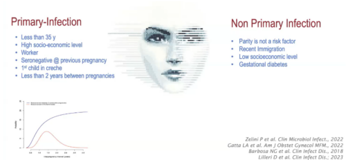
Prevention relies primarily on simple personal hygiene measures, including thorough handwashing after changing diapers or coming into contact with children’s saliva or urine, avoiding the sharing of eating utensils, and avoiding kissing young children on the mouth.
CMV vaccines are currently undergoing clinical investigation, but no vaccine has yet been approved for general use.
Seropositive women before and during early pregnancy
Pre-existing immunity to CMV does not provide complete protection, as viral reactivation or reinfection with a different CMV strain may occur.
Although current scientific evidence has not adequately established the effectiveness of hygiene measures in preventing non-primary CMV infection, adherence to these measures remains a reasonable preventive practice.
Diagnosis of Cytomegalovirus (CMV) Infection During Pregnancy
CMV infection in adults is usually asymptomatic, making clinical diagnosis difficult. The virus can be detected using molecular methods (PCR) or, less commonly nowadays, viral culture in biological specimens such as blood, urine, saliva, cervical secretions, and breast milk.
However, the detection of CMV DNA in a biological specimen does not, by itself, establish a recent primary infection.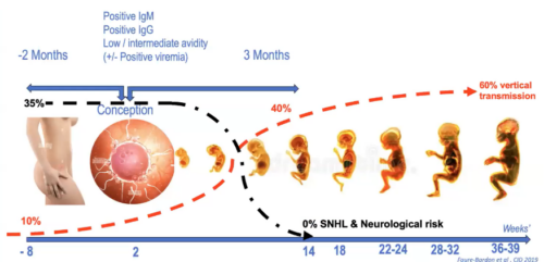
During pregnancy, diagnostic evaluation has three main objectives: identifying maternal infection, determining whether the infection is primary or non-primary, and investigating possible intrauterine transmission to the fetus.
Serological diagnosis: IgM and IgG antibodies
The initial laboratory investigation is based on the detection of CMV-specific IgM and IgG antibodies.
A positive IgM result alone does not establish a recent primary infection. IgM antibodies may persist for several months, reappear during viral reactivation or reinfection, or yield false-positive results due to cross-reactivity, including with other viral infections.
Similarly, negative IgM antibodies do not completely exclude a recent infection.
Documented IgG seroconversion, defined as the appearance of CMV-specific IgG antibodies in a woman with a previously documented negative result, is the most reliable serological evidence of primary infection.
An increase in IgG antibody levels in consecutive samples may support further investigation but is not, by itself, a reliable criterion for primary infection, particularly in the absence of a previously documented negative result.
In cases of borderline or discordant results, repeat testing is recommended, preferably using the same laboratory method or through a specialized reference laboratory.
CMV IgG avidity testing
IgG avidity testing evaluates the strength of binding between CMV-specific IgG antibodies and viral antigens, helping to estimate the timing of primary infection.
Low avidity: supports a recent primary infection, usually within the preceding 3–4 months, depending on the assay.
High avidity: particularly when detected during the first trimester, makes primary infection during the immediately preceding months less likely.
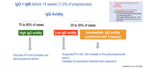
Intermediate avidity: does not allow reliable dating of infection and requires consideration of the clinical history, gestational age, previous serological results, and potentially repeat testing at a reference laboratory.
The interpretation of IgG avidity depends on the assay used and gestational age. Importantly, high avidity detected later in pregnancy does not exclude a primary infection during the first trimester.
Low avidity alone neither confirms fetal infection nor predicts its severity.
Special serological situations
In pregnant women with positive IgM and negative IgG, repeat serological testing after approximately two weeks is recommended to assess for possible IgG seroconversion.
CMV DNA detection by PCR in maternal blood may be used as an additional investigation in selected cases but does not replace serological assessment.
Diagnosing non-primary CMV infection is more challenging because IgG antibodies are already present, and no single serological marker can reliably distinguish viral reactivation from reinfection.
Furthermore, detection of CMV DNA in amniotic fluid confirms fetal infection but does not, by itself, distinguish primary from non-primary maternal infection.
Prenatal serological screening
Universal CMV serological screening during pregnancy remains subject to differing international recommendations and is not routinely recommended by several professional organizations.
Targeted investigation is appropriate when maternal infection is clinically suspected, exposure to CMV is considered possible, previous serological findings are suspicious, or fetal ultrasound abnormalities suggest congenital infection.
Repeated serological screening of seronegative pregnant women may be incorporated into specific antenatal surveillance protocols, although its frequency and duration are not universally standardized.
Prenatal diagnosis of fetal infection
In cases of confirmed or strongly suspected primary maternal CMV infection, or when suspicious fetal ultrasound findings are present, invasive prenatal diagnostic testing may be indicated.
Ultrasound findings suggestive of congenital CMV infection include ventriculomegaly, microcephaly, intracranial calcifications, abnormalities of brain development, echogenic bowel, hepatosplenomegaly, hepatic calcifications, ascites, fetal hydrops, and fetal growth restriction.
Amniocentesis followed by PCR detection of CMV DNA in amniotic fluid is the principal method for prenatal diagnosis of fetal CMV infection.
To minimize the risk of false-negative results, amniocentesis is generally performed after 20–21 weeks of gestation and at least 6–8 weeks after the estimated maternal infection. The precise timing depends on the clinical protocol.
A positive CMV PCR result in amniotic fluid confirms fetal infection but does not independently predict disease severity or the likelihood of long-term neurodevelopmental complications.
Prognostic assessment requires detailed specialist ultrasound examination, potentially fetal magnetic resonance imaging, and systematic follow-up.
Secondary Prevention of Congenital Cytomegalovirus (CMV) Infection
Secondary prevention aims to reduce the risk of intrauterine transmission of cytomegalovirus (CMV) from the mother to the fetus and, consequently, to prevent congenital infection.
In pregnant women with confirmed primary CMV infection during the periconceptional period or the first trimester of pregnancy, administration of high-dose valaciclovir, 8 g daily (2 g four times daily), has been associated with a significant reduction in transplacental viral transmission.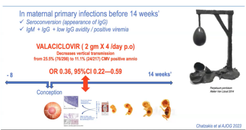
In a randomized clinical trial, valaciclovir treatment reduced the risk of fetal CMV infection by approximately 71% in relative terms, compared with placebo. This finding primarily concerns early primary maternal infection and should not be generalized to all cases of CMV infection during pregnancy.
Early initiation of treatment following diagnosis is considered important for its effectiveness, particularly when primary maternal infection occurs during the first trimester. The effectiveness of treatment for infections occurring later in pregnancy has not been established to the same extent.
Safety and adverse effects
Administration of valaciclovir at a dose of 8 g daily requires careful medical monitoring, as this dose is substantially higher than those commonly used for other herpesvirus infections.
Potential adverse effects include nausea, vomiting, and renal dysfunction, including acute kidney injury.
The original text reports gastrointestinal adverse effects in 21% of patients and mild-to-moderate acute kidney injury in 2%, with recovery following treatment discontinuation. These figures should be verified against the specific study from which they were derived before being presented as general risk estimates.
Adequate hydration, renal function monitoring, and dose adjustment when indicated are essential during treatment.
Clinical consideration: High-dose valaciclovir for the prevention of congenital CMV infection is not universally recommended by all international clinical guidelines. Treatment decisions should be individualized following specialist counselling and a careful assessment of potential benefits and risks.
Prenatal Diagnosis of Fetal CMV Infection
Prenatal diagnosis of fetal cytomegalovirus (CMV) infection is based on the detection of viral DNA in amniotic fluid using polymerase chain reaction (PCR) following amniocentesis.
Amniocentesis is generally performed after 20–21 weeks of gestation and at least 6–8 weeks after the estimated maternal infection, to minimize the risk of false-negative results.
Recent evidence suggests that, in selected cases and specialized centers, testing may be performed as early as 17 weeks of gestation, provided that at least eight weeks have elapsed since primary maternal infection. However, this approach requires appropriate counselling and consideration of its limitations.
A positive amniotic fluid PCR result confirms intrauterine infection but does not, by itself, establish fetal viremia or predict disease severity.
Negative amniotic fluid PCR
A negative PCR result, when amniocentesis has been performed at the appropriate time, substantially reduces the likelihood of fetal infection.
However, it does not completely exclude subsequent transplacental transmission or a false-negative result, particularly when testing is performed too soon after maternal infection.
The decision to discontinue preventive valaciclovir treatment following a negative PCR result should be individualized according to the treatment protocol, timing of maternal infection, and ultrasound findings.
Follow-up after confirmation of fetal infection
Once fetal CMV infection has been confirmed by positive amniotic fluid PCR, systematic surveillance at a specialized fetal medicine center is required.
Follow-up includes detailed ultrasound examinations every 3–4 weeks, with particular attention to fetal brain anatomy, growth, and extracerebral abnormalities.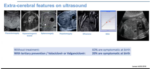
Fetal magnetic resonance imaging (MRI), usually performed between 28 and 32 weeks of gestation, provides additional assessment of the fetal brain, particularly when ultrasound findings are suspicious or inconclusive.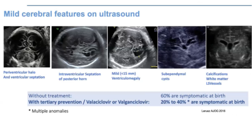
Repeat MRI may be considered in selected cases when additional prognostic information is expected.
The absence of significant abnormalities on detailed ultrasound examination and fetal MRI is a highly favorable prognostic finding. Nevertheless, it does not completely exclude sensorineural hearing loss or other delayed complications.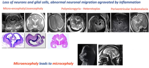
Fetal blood sampling to investigate thrombocytopenia has been used in selected specialist centers as an additional prognostic tool. However, because of the risks associated with invasive sampling, it is not routinely recommended.
Prenatal assessment of disease severity
Based on imaging and, when available, laboratory findings, fetal CMV infection may be broadly classified into three prognostic categories.
1. Fetal infection without detectable abnormalities
Amniotic fluid PCR is positive, but detailed ultrasound examination and fetal MRI reveal no significant abnormalities.
The overall prognosis is favorable, although sensorineural hearing loss remains possible. Hearing impairment may be unilateral or bilateral and may be present at birth or develop later.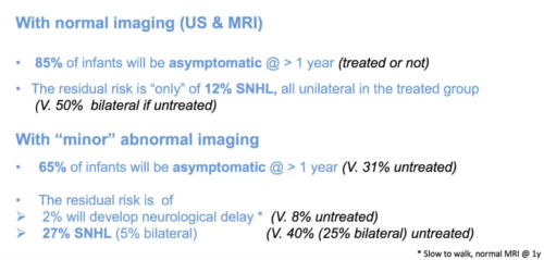
2. Mild-to-moderate symptomatic fetal infection
Limited abnormalities may be present, including echogenic bowel, hepatic calcifications, or mild ventriculomegaly.
Prognosis depends on the type, combination, and progression of these findings. Individualized counselling and systematic surveillance are required.
3. Severe symptomatic fetal infection
Severe infection is characterized by significant cerebral or systemic abnormalities, such as microcephaly, severe ventriculomegaly, cortical developmental abnormalities, extensive cerebral lesions, or fetal hydrops.
These findings are associated with an increased risk of adverse neurodevelopmental outcomes.
Multidisciplinary counselling is essential to discuss prognosis, available treatment options, continuation of pregnancy, and, where legally permitted, the option of pregnancy termination.
Clinical, Laboratory, and Imaging Findings of Congenital CMV Infection (cCMV)
The following findings are primarily assessed after birth, although several may also be identified prenatally.
Category | Clinical and diagnostic findings |
|---|---|
Clinical signs and symptoms | Intrauterine growth restriction or small size for gestational age, microcephaly, petechiae or purpura, blueberry muffin rash, jaundice, hepatomegaly, splenomegaly, lethargy, hypotonia, seizures, and feeding difficulties |
Laboratory abnormalities | Anemia, thrombocytopenia (for example, below 100,000/μL), leukopenia or neutropenia, elevated liver transaminases, and conjugated hyperbilirubinemia |
Cerebrospinal fluid (CSF) | Abnormal biochemical or cellular parameters and possible detection of CMV DNA |
Inflammatory or destructive brain lesions | Lenticulostriate vasculopathy, germinolytic pseudocysts, occipital horn septations, ventriculomegaly, periventricular calcifications, and white matter abnormalities |
Abnormalities of brain development | Polymicrogyria, abnormal cortical sulcation, and cerebellar hypoplasia |
Audiological findings | Unilateral or bilateral sensorineural hearing loss, which may develop or progress after birth |
Ophthalmological findings | Chorioretinitis or chorioretinal scarring |